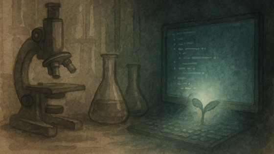
🔬 Up Close and Personal: Why Analyze in Color
 Rosalie Sinclair

Discover how dyes and microscopes reveal the hidden architecture of tomato roots, helping us grow smarter, more resilient crops from the ground up.
Some stories unfold underground. A tomato root stretches downward, carving its way through soil, quiet, steady, unseen. But under the microscope, the familiar soft blur of root becomes something dazzling. A landscape in miniature. Walls, seams, and swirling colors where cells once touched and traded the labor of growth.

To reveal this hidden world, scientists use a tool called a microscope, a device that magnifies tiny objects, letting us see details invisible to the naked eye. But there’s a catch: most plant tissues are nearly transparent, making it hard to distinguish one structure from another. That’s where dyes come in.
Toluidine blue stains this scene like watercolor. This special dye doesn’t just add color for beauty’s sake, it acts as a translator, turning the plant’s invisible
architecture into a vivid, readable map. Here’s how: Toluidine blue is what scientists call a “metachromatic” stain, meaning it reacts differently with various substances. When it meets lignin, a tough, woody polymer that strengthens certain cell walls, the dye turns those regions a striking pink or purple. Lignin is what gives the root its core strength, enabling it to push through the soil and withstand the elements. In contrast, areas rich in cellulose, a flexible, supportive
carbohydrate that forms the basic framework of plant cell walls, stay a cool blue. Cellulose is what allows the root to bend and grow.

The result is not only beautiful, but also informative. Each color tells a story about the root’s structure and function. The pink-stained regions mark the root’s reinforced core, while the blue zones reveal where growth and flexibility are needed.
Looking closer, you’ll see the root’s infrastructure in cross-section: xylem appears like pipelines, ready to carry water and minerals from the soil upward. Phloem forms highways, transporting sugars and nutrients to fuel the plant’s growth. These tissues, once invisible, now stand out in living color, thanks to the dye’s selective staining.
Even the tiniest details come into focus. Vesicles, tiny sacs inside the cells, move materials around, helping to build and maintain the cell walls. Every part, every color, has a role in the quiet complexity that makes a tomato, or any fruit, possible. What looks like art is a map, each color revealing the root’s structure and function.

Why do we go to such lengths? Because this kind of plant tissue analysis is more than just a window into beauty, it’s a toolkit for understanding and improving the crops that feed us.
By examining stained root slices, scientists can:
- Monitor Nutrient Status: The colors and patterns in the tissue reveal whether the plant is getting enough essential nutrients or if it’s suffering from hidden deficiencies that haven’t yet shown on the surface.
- Diagnose Problems Early: Before a plant shows outward signs of stress, tissue analysis can spot imbalances, allowing farmers to adjust fertilizer or water before yields are affected.
- Guide Precision Agriculture: With real-time information about what a plant needs, growers can fine-tune fertilizer and irrigation, reducing waste and protecting the environment from excess chemicals.
- Support Breeding and Research: Detailed tissue analysis helps plant breeders develop new varieties that are more resilient to disease, drought, or poor soils, by showing which genetic traits lead to stronger, healthier roots and stem.
- Track Plant Health and Disease: Scientists can use tissue staining to detect early signs of disease or stress, helping to breed or engineer plants with better resistance and to monitor the effectiveness of disease management strategies.
So, beneath the surface, this root is more than a structure; it’s a living record. The microscope and its dyes let us read that record, guiding decisions from the lab to the field, from the greenhouse to the dinner table. What we eat starts here, in the intimate, unsung architecture of a root, revealed in living color, and put to work for a more sustainable and abundant future.

Speaking of fruit, this season, tomatoes are making news not just underground, but in our gardens. A newly approved purple tomato, genetically engineered to boost antioxidant content, is now available to U.S. home gardeners. This vivid cultivar carries the same pigments that give blueberries their rich hue, and potentially, their health benefits. It’s a bold reminder that the color on your plate begins deep inside the plant, shaped by walls, genes, and time.

What we eat starts here, in the intimate, unsung architecture of a root. Thanks to the microscope and a splash of dye, we can finally see the story written in every cell.
This is the first in a new series exploring the structures that hold the plant world together. Stay tuned for pine needles, liverworts, and the vesicles that build walls from scratch.
References
Farmers Edge. (n.d.). 8 benefits of plant tissue testing. https://www.farmersedge.ca/8-benefits-of-plant-tissue-testing/
Megías, M. (n.d.). Techniques. Protocols TOLUIDINE BLUE. https://mmegias.webs.uvigo.es/02-english/5-tecnicas/02-toluidina.php
Motic Microscopes. (n.d.). Rapid preparation & examination of plant sections with microscopy. https://www.motic.com/asia/article-detail/rapid-preparation-and-examination-of-plant-sections-with-microscopy/
Penn State Extension. (n.d.). Plant tissue (total) analysis. https://extension.psu.edu/plant-tissue-total-analysis
Picketa Systems. (2023, October 18). Maximizing farm efficiency: The power of real-time plant tissue analysis. https://www.picketasystems.com/blog/maximizing-farm-efficiency-the-power-of-real-time-plant-tissue-analysis
ST Biologicals. (2023, May 1). How to read a plant tissue test. https://stbiologicals.com/how-to-read-a-plant-tissue-test/
STEM Learning. (n.d.). Microscopy of root tip mitosis. https://www.stem.org.uk/resources/community/collection/12852/microscopy-root-tip-mitosis
TPS Lab. (n.d.). The increasing need for plant tissue analysis. https://www.tpslab.com/the-increasing-need-for-plant-tissue-analysis
University of Maryland Extension. (n.d.). Plant tissue analysis. https://extension.umd.edu/resource/plant-tissue-analysis
Categories: : All, micro, plant structure